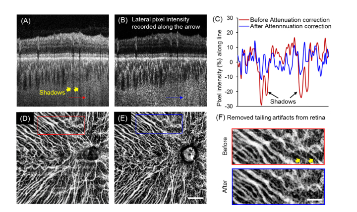
Fig. 4.
Attenuation correction eliminated shadows from retinal vessels and largely reduced the artifacts when visualizing the choroidal vasculature. (A-B) Representative B-scan before and after attenuation correction. Shadows from retinal vessels shown with yellow arrows were markedly reduced after attenuation correction. (C) Lateral pixel intensity profiles along the shadows (indicated with red and blue arrows) showed the percentage difference from the mean intensities before and after attenuation correction. After attenuation correction, the indicated shadows were successfully eliminated. (D-E) Minimum projection of choroidal vessels of a normal eye without (D) and with (E) attenuation correction. (Scale bar: 2 mm). (F) Magnified regions (red and blue squares) of the vasculature showed the elimination of artifacts from the retina after attenuation correction. (Scale bar: 1 mm).

