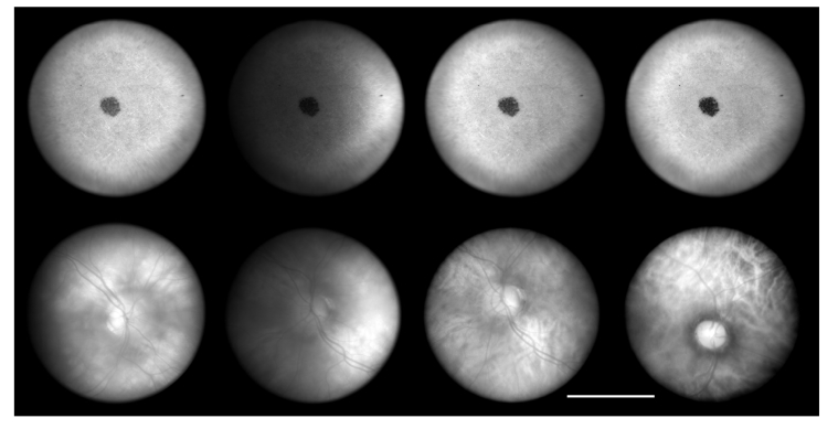
Fig. 13.
Images of a model eye (top) and two retinas (subject IDs ADS00054 and ADS00056), using 940, 850, 780 and 690 nm illumination (columns 1-4, respectively). Exposure times were 5, 10, 10 & 15ms, respectively. The black dot in the model eye was used to aid in alignment and focus. The scale bar is 15°.

