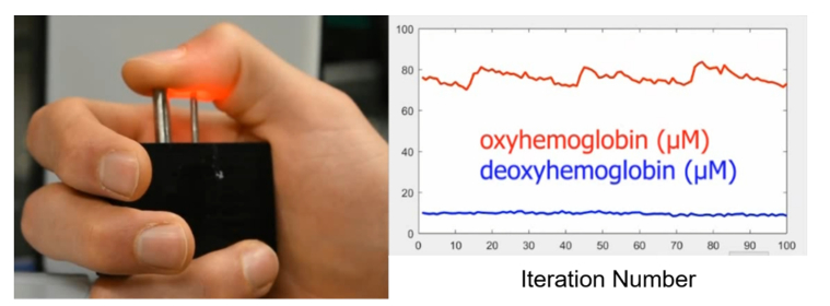
Fig. 3.
A still image from Visualization 1 (7.5MB, avi) depicting the instrument front-end showing real-time traces of oxy- and deoxy-hemoglobin during the measurement procedure. Inset: synchronized video of the subject’s hand during acquisition. Light from the source-fiber (right silver ferrule) passes through the thumb and is collected by the detector fiber (left silver ferrule).

