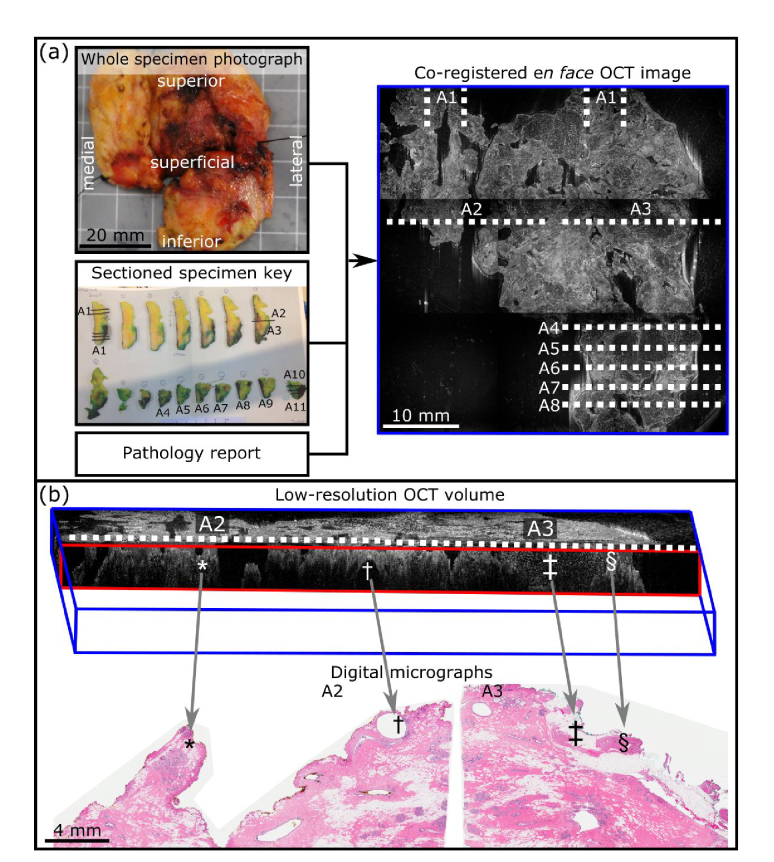
Fig. 2.
Histology co-registration and validation process. (a) Schematic outlining the process to estimate the location of tissue slices on the en face OCT image. (b) Validation of co-registration using low-resolution OCT volume to match digital micrographs. *, †, ‡, and § indicate co-registered features; *, change in surface topology; †, apocrine cyst; ‡, adipose tissue; § dense tissue.

