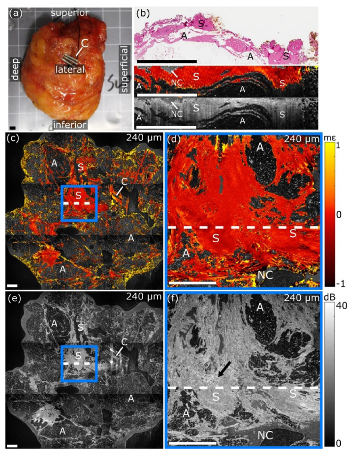
Fig. 3.
OCME of the lateral margin of freshly excised WLE specimen demonstrating contrast in uninvolved stroma (see Visualization 2 (8.8MB, avi) ). (a) Photograph of the lateral margin. (b) Digital micrograph and co-registered B-scans. (c) Wide-field and (d) magnified en face micro-elastogram. (e) Wide-field and (f) magnified en face OCT image. The arrow in (f) indicates an area of heterogeneous OCT intensity. En face images are presented at a depth of 240 µm. The white dashed lines in (c)-(f) indicate the location of the digital micrograph. All scale bars 3 mm. A, Adipose tissue; C, Clips; NC, Non-contact; S, Uninvolved stroma.

