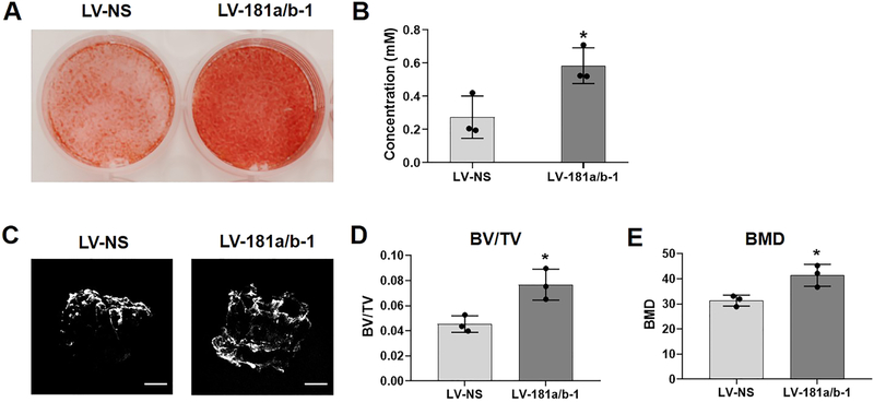
Fig. 2. Enhanced matrix mineralization by miR-181a/b-1.
Representative images of Alizarin red-stained cultures of osteogenic-induced DDCs transduced with either LV-NS or LV-181a/b-1 at day 14 (A). Quantification of Alizarin red staining is shown (B). Representative μCT images of human demineralized, decellularized bone scaffolds seeded with LV-NS or LV-181a/b-1-transduced cells and cultured in osteogenic medium for 28 days (C). Mineralization within the scaffolds was quantified by measuring bone volume / tissue volume (BV/TV) and bone mineral density (BMD) levels (D, E). Data in B, D, E expressed ± SD; n = 3. *p < 0.05. Scale bars in (C) = 1mm.

