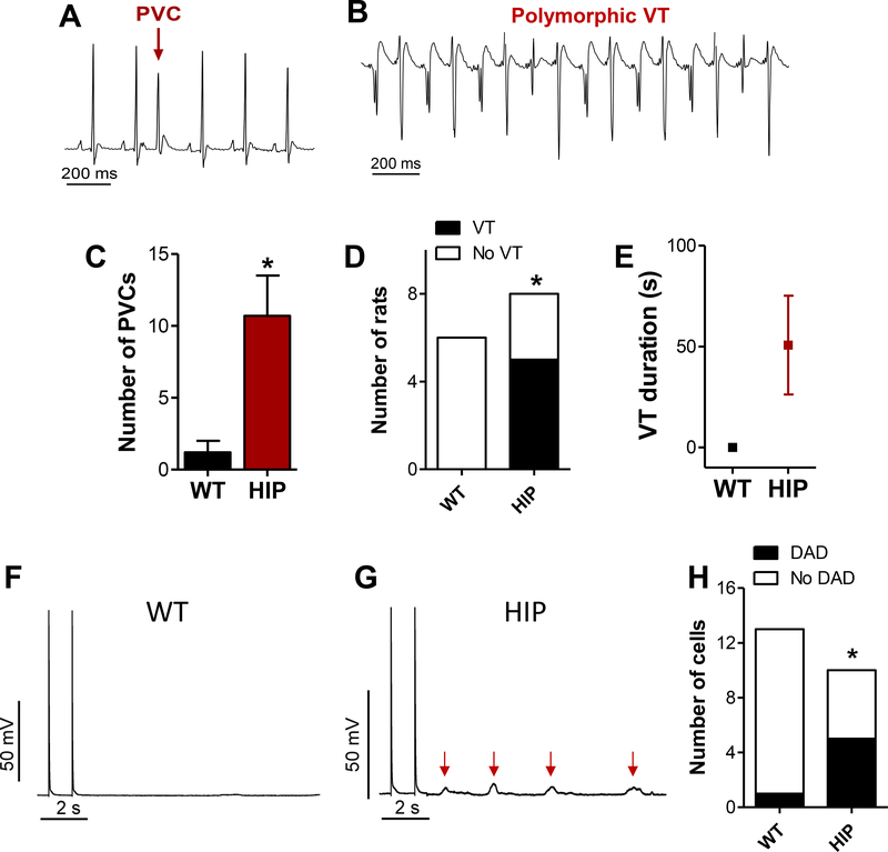
Figure 1. Abnormal electrical activity in diabetic HIP rats.
(A-B) Premature ventricular complex (PVC) and ventricular tachyarrhythmia (VT) recorded in HIP rats during a stress test with caffeine and dobutamine. (C) Mean number of PVCs recorded within 20 min following injection of caffeine and dobutamine in WT and HIP rats. (D) Number or rats showing VT within 20 min following injection of caffeine and dobutamine. (E) Average VT duration. (F-G) Measurement of DADs in WT (F) and HIP (G) myocytes. Current-clamped myocytes were pre-conditioned by pacing at 1Hz for 2 min. The occurrence of DADs was recorded for 10 s after pacing ended. Traces show the last two pacing-induced action potentials and the membrane potential during the following rest period. Red arrows point to DADs. (H) Frequency of DADs in myocytes from HIP and WT rats. N=13/10 myocytes/rats for WT and 15/10 for HIP. Statistical significance in panels D and H was assessed with Fisher’s exact test.

