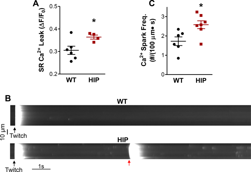
Figure 3. Higher SR Ca2+ leak and Ca2+ spark frequency in HIP rat myocytes.
(A) Total RyRmediated SR Ca2+ leak in WT and HIP myocytes. N=4 HIP and N=6 WT rats (3–8 myocytes/rat) (B) Representative line-scan recordings of spontaneous Ca2+ sparks in myocytes from HIP and WT rats preconditioned by steady-state pacing at 0.5 Hz (last stimulation-induced Ca2+ transient is indicated by black arrow). (C) Mean Ca2+ spark frequency in HIP and WT myocytes. N=7 HIP and N=6 WT rats (4–8 myocytes/rat). In (A) and (C), data were first averaged over cells from the same rat, then the mean SR leak/spark frequency per rat were averaged for each group.

