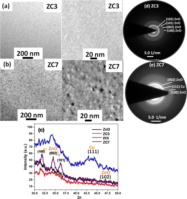
Figure 2.
TEM images [scale 200 nm (left) and 20 nm (right)] of Cu: ZnO thin film shows (a) untraceable Cu nano particle in ZC3 sample, (b) Cu nanoparticles (<10 nm) in ZC7 sample, (c) X-ray diffraction pattern of ZnO:Cu sample showing the crystalline nature of ZnO in low Cu concentration (≤5%) with no reflection of Cu and its oxide phases, and for high Cu concentration (≥7%) decrement of crystallinity of ZnO with broad reflection (at 2θ~43.4) of metallic Cu nano particle. TEM SAED pattern supports the XRD outcomes, (d) ZC3 sample represent the rings of crystalline ZnO, (e) ZC7 sample represent the ring of Cu alongwith ZnO.

