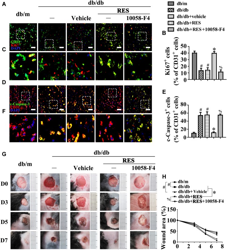
FIGURE 9.
c-Myc participates in the endothelial protective action of RES against hyperglycemia in vivo. (A) The presence of immunofluorescence with CD31 and Ki67 of wounded skin tissue sections, scale bars = 30 μm (×400), (C) enlargement of the indicated area in (A), scale bars = 12 μm (×1000), (D) confocal immunofluorescence with CD31 and c-Caspase-3 of wounded skin tissue sections, scale bars = 30 μm (×400), (F) enlargement of the indicated area in (D), scale bars = 12 μm (×1000), and (G) images of skin wounds, from db/m mice, db/db mice and db/db mice receiving RES (10 μM) or vehicle treatment with saline smeared on the wound, n = 6 mice in each group. For signaling pathway analysis, 10058-F4 (50 μM) was injected intradermally into the wound edges in the mice after RES smeared on the wound. Quantification of the proportion of Ki67 positive staining of endothelial cells (B), c-Caspase-3 positive staining of endothelial cells (E), and wound areas (H). All values displayed are means ± SEM of 8 independent experiments. #p < 0.05 vs. db/m mice; ∗p < 0.05 vs. db/db mice or vehicle treated db/db mice; %p < 0.05 vs. db/db mice receiving RES.

