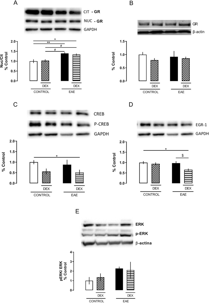
Figure 5.
Dexamethasone treatment increases cytoplasmatic GR and decreases EGR-1 expression, at the 8 dpi, in the EAE dorsal hippocampus of C57BL/6 female mice immunized with MOG35-55 plus CFA, treated (DEX + EAE) or not (EAE) with dexamethasone (DEX, 50 mg/Kg) on the day of immunization. Western Blot analysis of proteins in the cytosolic and nuclear fractions. Upper panel: Representative image of the Western blot assay. Bottom panel: densitometric analyzes of the specific bands represented in the upper panel. (A) Glucocorticoid receptor (GR) translocation expressed by the nuclear and cytosolic GR expression ratio. (B) Total expression of GR. (C) CREB activation expressed by the ration of the phosphorylated (pCREB) and total (CREB) forms of the transcription factor CREB in nuclear extracts. (D) Nuclear EGR-1 protein expression in nuclear extract. (E) ERK activation expressed by the ration of phosphorylated (pERK) and total (ERK) ERK cytosolic expression. The results are expressed as the mean ± SEM of the mean of the experimental groups. *p < 0.05 vs control; #p < 0.05 vs DEX; and $p < 0.05 vs EAE. Two-way ANOVA followed by Bonferroni post-test, n = 4 for each group.

