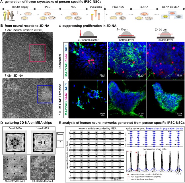
FIGURE 1.
Generation and analysis of synchronously active cortical 3D-neural aggregate cultures from human iPSC cultures. (A) Schematic representation of the in vitro neural induction protocol using dual SMAD inhibition to generate cortical 3D-neural aggregate cultures. (B) Phase-contrast images show an overview of cell morphology in hiPSC-NSC cultures at 1 and 7 days after seeding of hiPSC-NSC cryovials. Rectangles show the transition from neural rosette (red rectangle) toward 3D-neural aggregate (blue rectangle) within 7 days in vitro (div). (C) Confocal images visualize MAP2-AB+-neurons and Ki-67+-cells in neural rosette and 3D-neural aggregates (day 30 post iPSC stage) under the indicated culture conditions. The schematic drawings illustrate the regions and z-levels of image acquisition. (D) Three different donor-derived hiPSC-NSC lines were used to generate 3D-neural aggregates seeded on 6-well (9 electrodes/well) or single well (60 electrodes/well) MEAs. (E) After MEA recordings, spikes were detected by an automatic spike detection algorithm and extracted from raw data by the Spanner software. Spike data were used for population bursts detection [custom-made Matlab tools as described in (Hedrich et al., 2014)].

