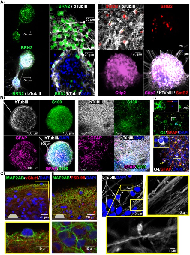
FIGURE 6.

Confocal images show cortical neuronal identity, presence of glial cells, synapses and spines in 3D-neural aggregate cultures. (A,i) BRN2+, (A,ii) CTIP2+, and SATB2+ cortical βTubIII+-neurons within 3D-neural aggregates. (B,i,ii) βTubIII+-postmitotic neurons were embedded in a glial network comprising GFAP+, S100β+ astrocytes and (B,iii) O4+ oligodendrocytes present outside (above) and at the edges (below, arrows) of 3D-neural aggregates. Boxes show morphology of O4+ oligodendrocytes at higher magnification. (C,i,ii) Visualization of excitatory vGlut+ and PSD-95+ synapses as well as (C,iii) βTubIII+-filled spines within 3D-neural aggregates. All images were taken 28 days after neural aggregate isolation and cultivation of ChiPSC4-derived neural cultures.
