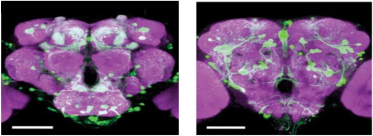
FIGURE 2.
DA cells in the Drosophila brain. An anterior view of DA neurons in the Drosophila brain. Labeling of DA cells and processes was achieved by a tyrosine-hydroxylase enhancer trap driving expression of green fluorescent protein (White et al., 2010), Scale bar, 100 μm (courtesy of Dr. Frank Hirth, King’s College London).

