Figure 1.
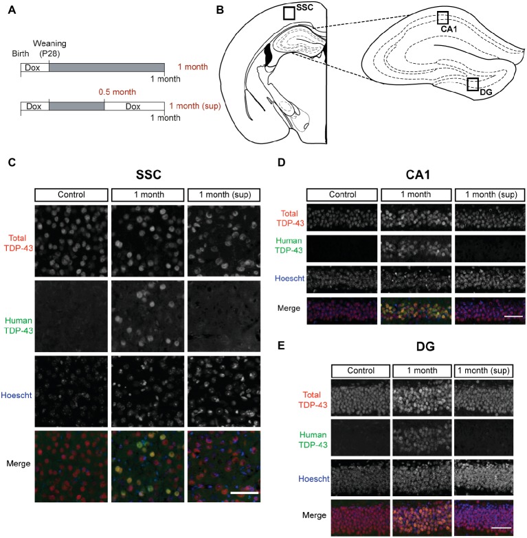
TDP-43 expression pattern in inducible TDP-43-WT transgenic mice. (A) Experimental design: transgene expression was inactive until postnatal day 28 by treatment with Dox. For the suppression protocol, mice were treated again with Dox at 0.5 month post-weaning to suppress transgene expression for 2 weeks. The behavioral analysis (motor, cognitive, and social) was performed at 0.5 month post-weaning (0.5 month mice) and at 1 month post-weaning [1 month (sup) mice]. The results on these mice were compared with mice in which transgene expression was preserved until 1 month post-weaning (1 month mice). (B–E) Expression of human TAR DNA-binding protein 43 (TDP-43) in tTA/WT12 mice. (B) Schematic diagrams (adapted from Paxinos and Franklin, 2008) showing different hTDP-43-expressing brain areas (indicated by the labeled boxes) such as somatosensory cortex (SSC), hippocampal cornus ammonis 1 (CA1), and dentate gyrus (DG). (C–E) Double immunofluorescence of total TDP-43 (human + mouse TDP-43) and human TDP-43 in representative coronal brain sections of control, tTA/WT12 1 month and tTA/WT12 1 month (sup) mice. High power micrographs of boxed areas in B are shown: SSC (C), hippocampal CA1 region (D), and DG (E). Scale bar: 50 μm.

