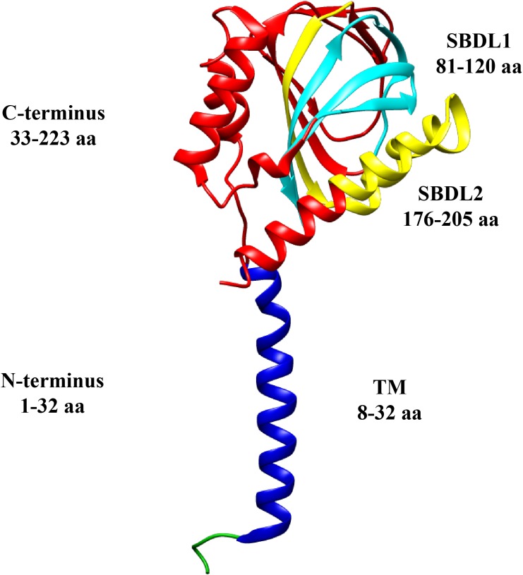
FIGURE 1.
Sigma-1 receptor topology. Structural domains of a monomer of the Sig-1R are shown in different colors: N-terminus (green and blue), transmembrane segment (TM, blue), C-terminus (red), and the two Steroid Binding Domain-Like (SBDL1, aqua, and SBDL2, yellow) which are located in the C-terminus. The amino acids (aa) comprising each domain are illustrated (PDB structure entry, 5HK).

