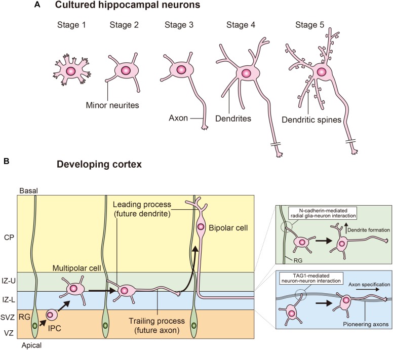
FIGURE 1.
Processes of neuronal polarization in vitro and in vivo. (A) Upon isolation, hippocampal neurons form filopodia (stage 1). These neurons subsequently extend several minor neurites (stage 2), which show characteristic alternations of growth, and retraction. The major polarity event occurs when one of these equivalent minor neurites grows rapidly to become the axon (stage 3). The next steps are the morphological development of the remaining short minor neurites into dendrites (stage 4) and the functional polarization of axons and dendrites, including dendritic spine formation (stage 5). (B) In the developing neocortex, cortical neurons are generated from the radial glia in the VZ. These neurons extend multiple minor neurites and migrate toward the IZ through the SVZ. The multipolar (MP) cells generate a trailing process (future axon) and a leading process (future dendrite), and then these MP cells transform into bipolar (BP) cells and migrate toward the cortical plate (CP). A TAG-mediated interaction between a minor neurite of an MP cell and pioneering axons determines axon specification in the lower part of the IZ (IZ-L). N-cadherin-mediated interactions between the radial glia and neuron interactions determines dendrite specification in the upper part of the IZ (IZ-U).

