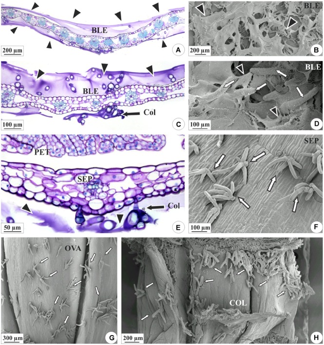
FIGURE 2.

Distribution of the colleters (arrows) and secretion in the floral organs of E. brasiliensis. (A,C,E) Light microscopy. (B,D,F–H) Scanning electron microscopy. (A–D) Bracteole covered by a large amount of secretion (arrowheads). (C,D) Magnified detail of the colleters (“Col” and white arrows) involved in secretion. (E,F) Sepals presenting colleters and a lower amount of secretion. (G) Ovarian wall showing colleters. (H) Floral column with colleters on the surface. BLE, bracteole; COL, floral column; OVA, ovary; PET, petal; SEP, sepal.
