
FIGURE 3.
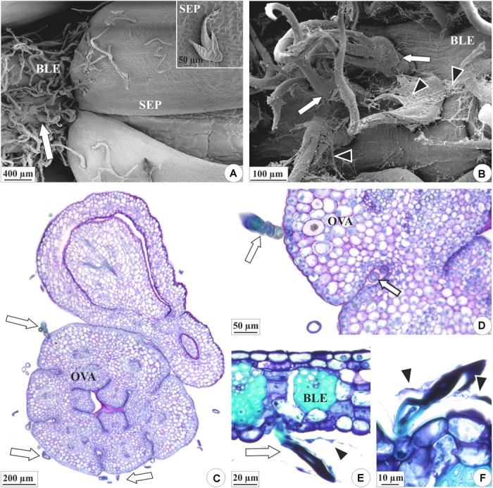
Distribution of the colleters (arrows) and secretion in the floral organs of E. crinipes. (A,B) Scanning electron microscopy. (C–F) Light microscopy. (A) Floral bud with bracteole showing colleters (white arrow) and sepals surrounding the bud. Inset: colleter on the surface of the sepal. (B) Magnified detail of the bracteole, showing colleters, and secretion (arrowheads). (C) Ovary with colleters on outer surface, as viewed from above the ovary axis of a lateral flower surrounded by a bract. (D) Detail of the ovary, showing colleter on the surface and the recess of the wall (white arrow). (E) Bracteole with a colleter. (F) Detail of the bracteole’s colleters surrounded by their secretion. BLE, bracteole; OVA, ovary; SEP, sepal.