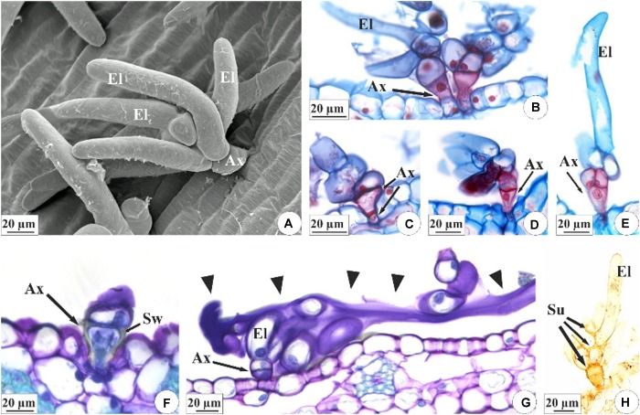
FIGURE 4.
Characterization of the colleters of E. brasiliensis (A–E,G,H) and E. crinipes (F). (A) Scanning electron microscopy. (B–H) Light microscopy. (A) Brush-type colleter with short basal axis and elongated terminal cells. (B–E) Colleter with short cup-shaped basal axis composed of one (B), two (C,D), or three cells (E), and presenting lignified and suberified secondary wall deposition, as indicated by double-staining with afstra blue and safranin. Elongated cells have only primary cell walls. (F) Detail of the basal axis of the colleter showing the cup-shaped cell with a secondary wall. (G) Colleter with elongated cells releasing secretion (arrowheads) that covers the surface of the bracteole. (H) Histochemical test with Sudan IV indicating suberification in the cells of the colleter’s axis and in the cell walls of the elongated cells that connect with the cells of the axis. Ax, axis of colleter; El, elongated cell; Sw, secondary wall; Su, suberized wall.

