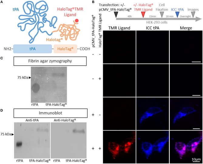
Figure 1.
Generation of the tPA-HaloTag®. (A) Scheme of the tPA-HaloTag® protein: tPA in blue (69 kDa), HaloTag® in orange (33 kDa) and the specific fluorescent exogenous ligand (HaloTag® TMR Ligand) in red. (B) Representative confocal images of HEK-293 cells transfected (+) or not (–) with pCMV_tPA-HaloTag®, incubated 48 h later (+) or not (–) in the presence of the HaloTag® TMR ligand, followed by immunocytochemistry (ICC) raised against tPA (blue). Scale Bar: 10 μm. (C) Fibrin–agar zymography showing the fibrinolytic activity of recombinant tPA (rtPA) as control and tPA-HaloTag® produced by HEK 293 transfected cells. (D) Immunoblots of recombinant tPA (rtPA) and proteins from HEK-293 cells culture transfected with pCMV_tPA-HaloTag® revealed with antibodies raised against either tPA or HaloTag®. Control tPA (rtPA).

