Figure 5.
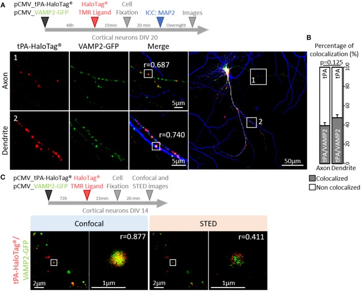
tPA is associated with VAMP2 proteins. (A) Timeline of the experiments. Representative z-stack images of co-transfected cortical neurons (at DIV20) with pCMV_tPA-HaloTag® (TMR ligand, in red) and pCMV VAMP2-GFP (in green), followed by ICC raised against MAP2 (in blue). tPA-HaloTag® colocalizes with VAMP2-GFP (exosome) in axons (MAP2-; frame 1) and in dendrites (MAP2+; frame 2) with Pearson's coefficients, respectively: r = 0.687 and r = 0.740. Scale Bar: 5 or 50μm (whole neuron). (B) Representative diagrams of the percentages of colocalization between tPA-HaloTag® and VAMP2-GFP in function of total tPA-HaloTag® puncta. There are 39% of colocalization in axons and 48% in dendrites (p = 0.125). (C) Representative images of co-transfected cortical neurons cortical neurons (at DIV14) with pCMV_tPA-HaloTag® (reveals by TMR ligand, in red) and pCMV_VAMP2-GFP (in green). tPA-HaloTag® and VAMP2-GFP colocalize, when revealed by confocal and super-resolution microscopy: Stimulated Emission Depletion (STED) with Pearson's coefficients, respectively: r = 0.877 and r = 0.411. Scale Bar: 2 or 1 μm.

