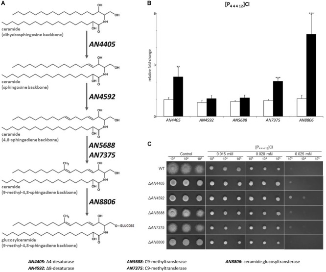
FIGURE 3.

(A) Schematic representation of the glucosylceramide pathway. (B) Expression analysis of glucosylceramide pathway genes after 4 h of exposure to dodecyltributylphosphonium chloride ([P44412]Cl, black bars), compared to the control (white bars). Axis y represents the fold-change relative to time-zero (∗∗p < 0.01, ∗∗∗p < 0.001). (C) Growth assessment in solid media supplemented with 0.015, 0.020, or 0.025 mM of [P44412]Cl. 105, 104, and 103 conidia were inoculated in each plate.
