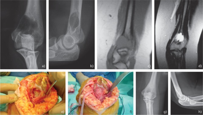
Fig. 2.
(a) Anteroposterior and (b) lateral radiographs of the elbow showing an aneurysmal bone cyst of the distal humerus in 17-year-old female. (c) Τ1 and (d) T2 magnetic resonance images of the elbow. (e) Intraoperative images showing the gross destruction of the distal humerus. (f) The lesion was treated with curettage and bone grafting. (g) Anteroposterior and (h) lateral radiographs of the elbow 11 years postoperative showing a good incorporation of the graft and no sign of recurrence.

