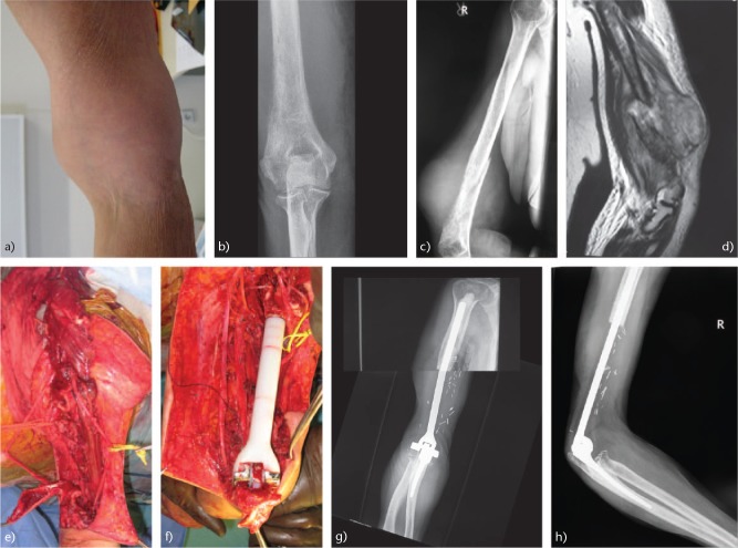
Fig. 3.
(a) A rapidly enlarging mass in the right arm of a 69-year-old female. (b) Anteroposterior radiograph of the distal humerus showing a lytic lesion with permeation of lateral cortex. (c) High-grade sarcoma was diagnosed. Pathological fracture of the distal humerus. (d) T1-MRI image showing the tumour mass. (e) Intraoperative image of the right humerus after excision of the tumour with preservation of the neurovascular elements. (f) Elbow reconstruction using a custom-made cemented megaprosthesis (Link megaprostheses, Hamburg, Germany). (g) Anteroposterior and (h) lateral radiographs of the elbow 13 months postoperatively, showing the elbow endoprosthesis with no sign of local recurrence. Postoperatively, the patient had adjuvant chemotherapy. She died at 13 months due to lung metastatic disease.

