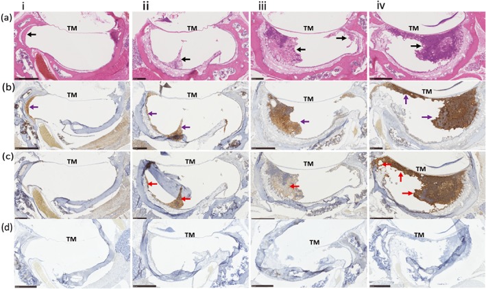
Figure 4.

Immunostaining of NTHi inoculated Junbo mouse middle ears. Formalin fixed paraffin embedded sections of four NTHi inoculated Junbo mouse middle ears (i,ii,iii,iv) with variable volumes and grades of ear fluid (a) Haematoxylin and Eosin‐stained section of middle‐ear bulla, showing the tympanic membrane and the middle‐ear fluid—black arrow. (b) DAB immunostaining of neutrophils in the middle‐ear fluid with rabbit anti‐MPO antibody showing presence in the fluid—purple arrow. (c) DAB immunostaining of NTHi 162 sr in the middle ear fluid with anti‐NTHi162 antibody showing the presence and uneven distribution of bacteria across the fluid—ense patch indicated by red arrow. (d) Staining using control rabbit serum (non‐immune) as primary antibody and rabbit VERTEX ABC kit for detection
