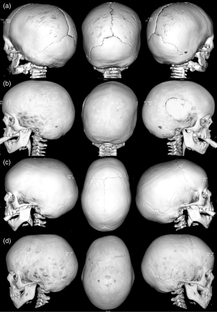
Figure 3.

Selected 3D‐CT scan views from two probands illustrating the progressive nature of the craniosynostosis. (a) and (b) Patient 14 (K3): 3D‐CT images taken at age 0.8 and 2.7 years, respectively. At 0.8 years only the squamosal sutures were noted to be closed, progressing to pansynostosis with associated papilledema by 2.7 years. The site of the neurosurgical evacuation of a presumed spontaneous extradural bleed is also visible. Note the relatively normal skull shape. (c) and (d) Patient 8 (K3): 3D‐CT images taken at ages 1.9 and 4.7 years, respectively. At 1.9 years there was a scaphocephalic head shape with an indistinct sagittal suture suspicious of synostosis. By 4.7 years when clinical evidence of raised intracranial pressure became apparent, the craniosynostosis had progressed with clear involvement of the sagittal, superior bilambdoid, left inferior coronal, and left squamosal sutures
