Figure 3.

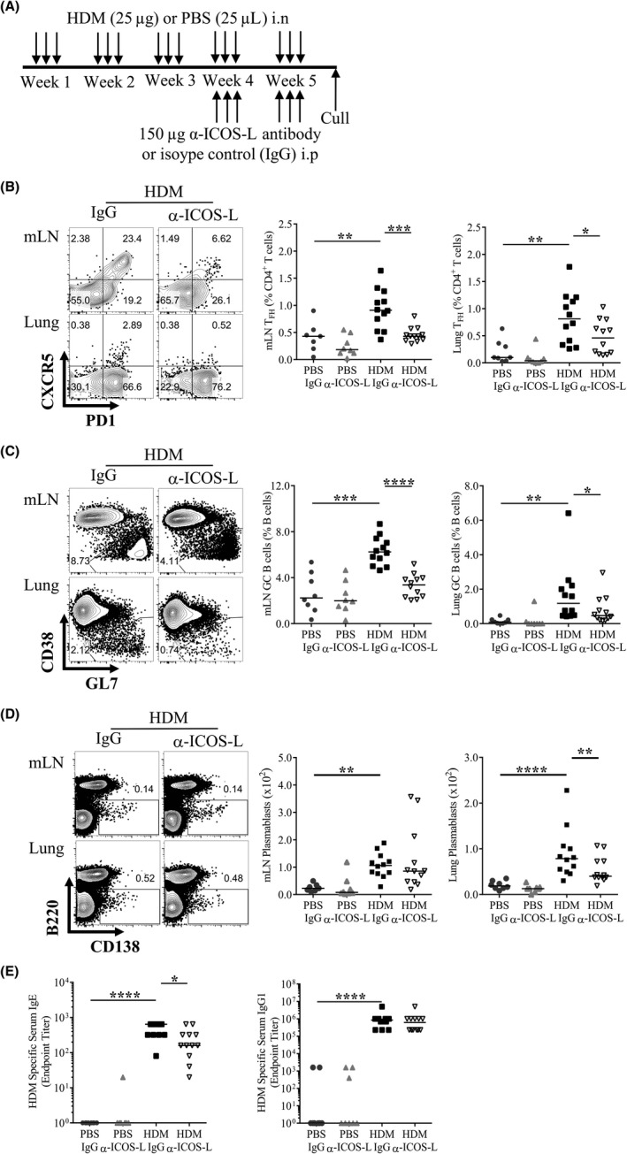
ICOS/ICOS‐L interactions are required to sustain TFH during chronic allergic airway disease (AAD). Adult female BALB/c mice were exposed (i.n) to 25 μg house dust mite (HDM) or 25 μL phosphate‐buffered saline (PBS), three times a week for 5 weeks. From the start of week 4, mice were also administered 150 μg anti‐ICOS‐L (α‐ICOS‐L) or isotype control (IgG) antibody (i.p) three times a week. Mice were culled at the end of week 5. A, Schematic of experimental design, B, Representative flow plots of mLN and lung TFH following HDM and IgG or α‐ICOS‐L treatment. C, Representative flow plots of mLN and lung germinal centre (GC) B cells following HDM and IgG or α‐ICOS‐L treatment. Pre‐gated on CD19+B220+ B cells. The data are quantified for all groups. D, Representative flow plots of mLN and lung B220− CD138+ plasmablasts following HDM and IgG or α‐ICOS‐L treatment, pre‐gated on lymphocytes. Plasmablast numbers are quantified for all groups. E, Serum was titrated, and allergen‐specific antibody was measured by ELISA. Endpoint titres are displayed for IgE and IgG1. Statistical significance was determined using a Mann‐Whitney U test. *P < 0.05, **P < 0.01, ***P < 0.001. Data are pooled from two independent experiments, n = 8 for PBS‐treated groups, n = 12 for HDM‐treated groups
