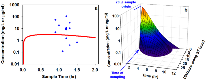
Figure 8:
(a) Comparison of the dexamethasone time course from a simulation representing the human (red line) with perilymph sample measurements from humans as reported by Bird et al., 2011 (blue symbols). The simulation used kinetic parameters derived from the guinea pig data in this study and inner ear dimensions corresponding to the human. The model prediction lies well within the scatter of measurements, validating the ability of the model with the parameters derived here to predict dexamethasone concentrations within the range of those measured in the human cochlea. (b) Distribution of dexamethasone as a function of distance and time along scala tympani from the same simulation in which the line in (a) was derived. Intratympanic dexamethasone-phosphate therapy is calculated to provide only a brief exposure of basal half of the cochlear to drug. The spatial origin and the timing of the 20 μL samples collected by Bird et al. are indicated.

