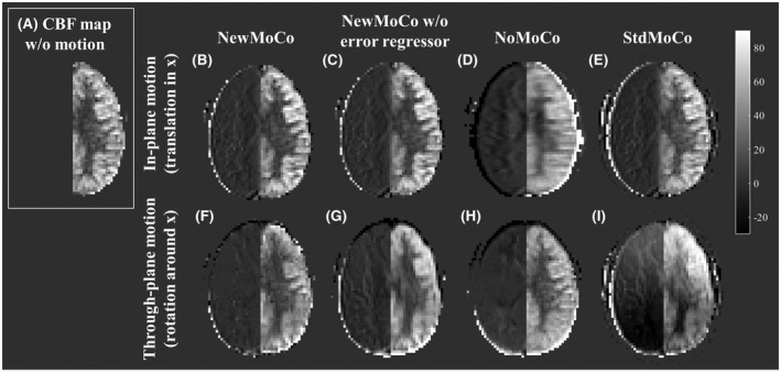
Figure 5.

Representative CBF maps obtained from simulation 2. In the right hemisphere, no perfusion signal was simulated, and therefore all signal intensity in the right hemisphere indicates motion‐related errors. (A) Reference CBF map obtained from the data set without motion. (B–E) Respectively NewMoCo, NewMoCo without error regressor, NoMoCo, and StdMoCo are applied to in‐plane motion (translation in the x‐direction) and (F–I) to through‐plane motion (rotation around the x‐axis)
