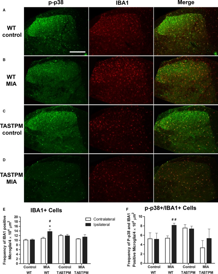
Figure 5.

Lack of MIA‐induced spinal microgliosis exhibited by TASTPM mice. Representative images showing phospho‐p38 (p‐p38) expressing microglia (IBA1) in the dorsal horn of the spinal cord (L4–L5) 28 days after intra‐articular injection of saline (control) or MIA of TASTPM and age‐ and gender‐matched wild‐type (WT) mice (A–D). The scale bar represents 200 μm. Quantification of IBA1 immunopositive cells (E) and P‐p38 expressing IBA1 cell frequency (F) was conducted in pooled L4 and L5 dorsal horn of MIA and control mice. WT mice exhibited significantly greater frequency of IBA1 immunopositive microglia in the ipsilateral dorsal horn compared to WT control ipsilateral (*p < 0.05, Student's t‐test) and WT MIA contralateral, where increase in activated microglia was also evident (# p < 0.05, ## p < 0.01, Student's t‐test). Contrastingly, no apparent changes in microglial or activated microglia were observable in the ipsilateral MIA‐injected TASTPM mice compared to neither TASTPM control ipsilateral nor TASTPM MIA contralateral (p > 0.05, Student's t‐test). Data are shown as mean ± SEM (n = 4 (2 males and 2 females) per experimental group).
