
Figure 1.
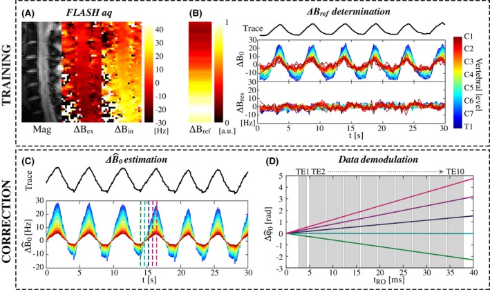
Schematic of the trace‐based correction method. A, Magnitude and phase images of a single sagittal slice are acquired with a FLASH sequence (data shown for subject 4). The phase images are here shown at a peak of expiration () and inspiration (). B, The coupling parameter () between the respiratory trace and the field offset () inside the spinal cord is determined for each axial slice based on the FLASH calibration data. The residual field offsets () show that the linear model explains a large part of the measured temporal field variations. C, During later scans, the field offset () can be estimated based on and the respiratory trace. The color scale to indicate vertebral level is the same as in B. D, The estimated field offset yields corresponding phase evolutions (), here shown for a multi‐echo gradient‐echo readout at 5 different time points indicated by vertical lines in the plot in C. The acquired image data are demodulated by the estimated