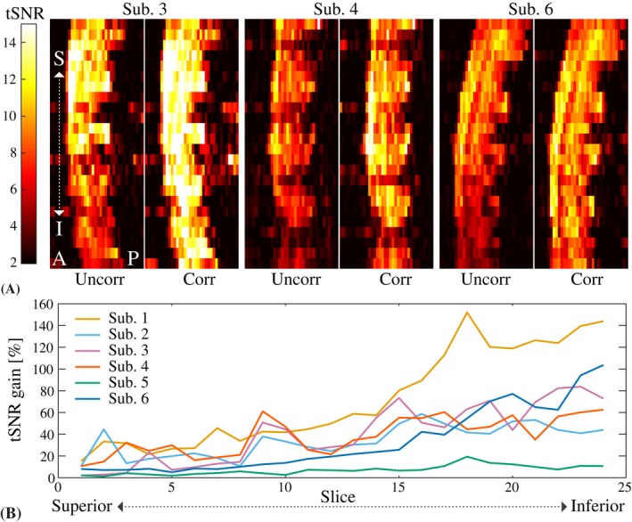
Figure 5.

A, Sagittal tSNR maps for the multi‐shot EPI without and with correction in 3 different subjects. B, Relative tSNR gain due to the correction for all subjects. The tSNR was averaged over all voxels inside the spinal cord mask in each axial slice (slice numbers counted superior to inferior, covering approximately C2 to C6)
