Figure 1.

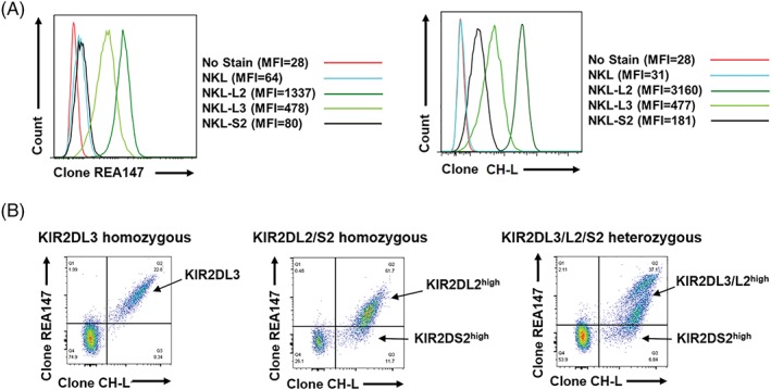
Discrimination of KIR2DS2 from KIR2DL2 and KIR2DL3. A, NKL cell lines either untransfected or transfected with KIR2DL2, KIR2DL3 or KIR2DS2 were stained with antibody clone REA147‐FITC at 1:10 dilution (Miltenyi Biotech) or antibody clone CH‐L‐PE at 4:100 dilution (BD Biosciences) and analysed by flow cytometry. B, Representative flow cytometry plots of primary human CD3‐CD56+ natural killer (NK) cells stained with REA147 and CH‐L are shown from KIR2DL3 homozygous (of seven donors), KIR2DL2/KIR2DS2 homozygous (of four donors) and KIR2DL3/KIR2DL2/KIR2DS2 heterozygous (of 10 donors) donors (assessed by PCR using sequence specific primers18). The KIR2DL3/L2high and KIR2DS2high NK cell populations detected by flow cytometry are indicated
