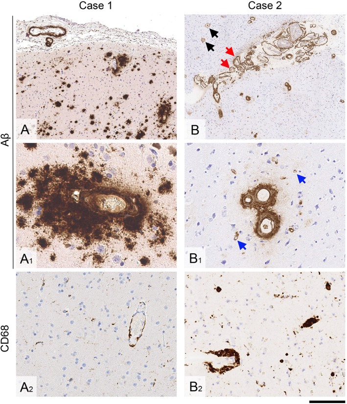
Figure 2.

Brain biopsy findings. (A–A2) In Case 1, there is leptomeningeal and cortical cerebral amyloid angiopathy, and widespread diffuse parenchymal amyloid‐β (Aβ) deposits (A and A1). There is only minimal parenchymal and perivascular microglial and macrophage activity (A2). (B–B2) In Case 2, there is particularly widespread leptomeningeal and cortical amyloid angiopathy, including vessel wall splitting (B, red arrows) and capillary amyloid angiopathy in the cortex (B1, blue arrows). In the cortex, there are also diffuse parenchymal Aβ deposits and rare plaques with central amyloid cores (B, black arrows). There are abundant perivascular macrophages, some laden with hemosiderin (B2). Sections A, A1, B, and B1 are immunostained for Aβ; A2 and B2 are immunostained for CD68. All sections are counterstained with hematoxylin. Scale bar = 500 μm in A and B; 50 μm in A1; 100 μm in B1, and 200 μm in A2 and B2. [Color figure can be viewed at www.annalsofneurology.org]
