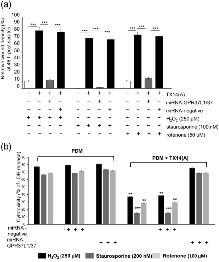
Figure 5.

TX14(A) acts via GPR37L1 and GPR37 to protect primary astrocytes against toxicity induced by H2O2, staurosporine or rotenone. (a) Pre‐exposure to stressors (5 hr) drastically reduce relative wound density recorded at 48 hr in PDM. TX14(A) (100 nM) rescued astrocytes bringing wound density close to normal (compare to Figure 3). GPR37L1/GPR37 knock‐down prevented the protective effect of TX14(A), while the control vector had no effect (n = 6, triplicates, ***p < .001 vs. indicated group). (b) LDH release was used as a measure of cytotoxicity, 24 hr after exposure of astrocytes to oxidative stress. In PDM, manipulation of GPR37L1/GPR37 had no effect. TX14(A) (100 nM) protected them from damage but only when they were expressing GPR37L1/GPR37, (n = 6, triplicates, **p < .01, ***p < .001 vs. control group, for example, PDM groups). One‐way ANOVA with Bonferroni's post hoc analysis [Color figure can be viewed at wileyonlinelibrary.com]
