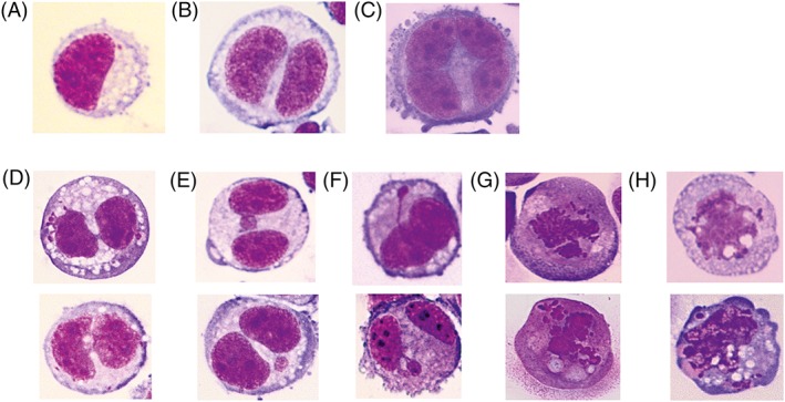
Figure 2.

Photographic images of the cells and different nuclear anomalies (NBs – nucleoplasmic briges; NBuds – nucleoplasmic buds), stain Diff Quick, magnification 400×, (A) mononucleated epithelioid cell with some cytoplasmic vacuoles and few desmosomes; (B) binucleated epithelioid cell; (C) multinucleated epithelioid cell with desmosomes; (D) binucleated epithelioid cells with some cytoplasmic vacuoles and one or two nucleoplasmic bridges; (E) binucleated epithelioid cells with a micronucleus in different position; (F) binucleated epithelioid cells with nuclear buds; (G) mononucleated apoptotic cells; (H) mononucleated necrotic cells.
