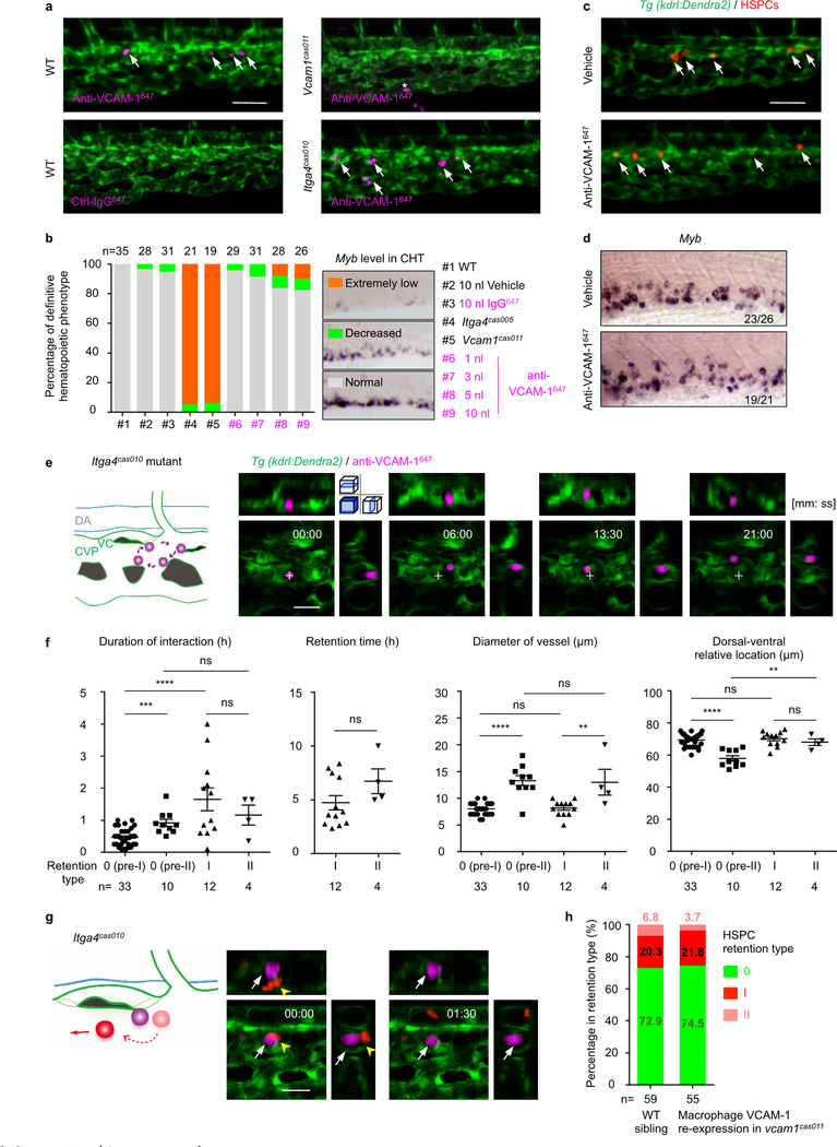
Extended Data Fig. 9 |. Anti-VCAM-1647 antibody labels usher cells without disrupting definitive haematopoiesis.
a, Injection of 1 nl (0.4 ng) of anti-VCAM-1647 antibody labels usher cells (arrows) in wildtype and itga4cas010 mutants in the Tg(kdrl:eGFP) background, whereas injection of either control (non-specific) IgG647 antibody into wildtype cells or anti-VCAM-1647 antibody into vcam1cas011 mutants in the Tg(kdrl:eGFP) background did not label any cells in the retention hotspots. Asterisk indicates nonspecific labelling on a chromatophore in the CHT. b, Anti-VCAM-1647 injection marginally influence definitive haematopoiesis. Statistical analysis shows the percentage of the three types of definitive haematopoietic phenotype in nine different conditions, including wild-type embryos without injection (#1), with 10 nl vehicle (#2) or 10 nl 0.4 mg ml−1 IgG647 injection (#3), itga4cas005 mutants (#4) or vcam1cas011 mutants (#5) without injection, and wild-type embryos with 1–10 nl 0.4 mg ml−1 anti-VCAM-1647 injection (#6-#9). c, d, Live imaging of HSPCs (c) or WISH analysis of the myb probe at 60 h.p.f. (d) of the wild-type CHT after vehicle or 1 nl anti-VCAM-1647 antibody injection. e, Schematic diagrams (left) and confocal imaging (right) show VCAM-1+ cells patrolling on a small scale in the CHT in itga4cas010 mutant embryos. Cross indicates the original position at the initial time point. f, Statistical analysis of the duration of the interaction between HSPCs and usher cells, the HSPC retention time, the diameter of vessels and the dorsal-ventral relative location for HSPC retention in pre-type I (type 0), pre-type II (type 0), type I and type II. Duration, pre-I vs pre-II: ***P = 0.0001, t = 4.25, df = 41; duration, pre-I vs I: ****P< 0.0001, t = 5.31, df = 43; duration, pre-II vs II: P = 0.37, t = 0.93, df = 12; duration, I vs II: P = 0.46, t = 0.76, df = 14; retention: P = 0.16, t = 1.50, df = 14; diameter, pre-I vs pre-II: ****P < 0.0001, t = 8.80, df = 41; diameter, pre-I vs I: P = 0.68, t = 0.42, df = 43; diameter, pre-II vs II: P = 0.89, t = 0.14, df = 12; diameter, I vs II: **P = 0.006, t = 3.24, df = 14; location, pre-I vs pre-II: ****P < 0.0001, t = 7.64, df = 41; location, pre-I vs I: P = 0.6, t = 0.53, df = 43; location, pre-II vs II: **P = 0.005, t = 3.41, df = 12; location, I vs II: P = 0.41, t = 0.85, df = 14. g, In itga4cas010 mutants, HSPCs encountered but failed to interact with usher cells and then went through the CHT within a few minutes (see Supplementary Video 10). h, The percentage of the type 0, type I and type II HSPC retention types in wild-type sibling and vcam1cas011 mutants in the Tg(mpeg1:Gal4,kdrl:Dendra2) background with transient transgenesis of UAS:vcam1. None of the HSPCs in the vcam1 mutants could be classified into either type I or II retention types, or were comparable with the HSPCs in Extended Data Fig. 8h. Scale bars, 50 μm (a, c) and 20 μm (e, g).

