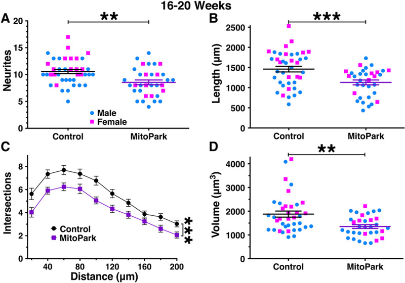
FIG. 3.

Morphological parameters are mildly reduced in dopamine neurons from 16- to 20-week-old MitoPark mice. By 16 to 20 weeks of age, dopamine neurons from male (blue circles) and female (pink squares) MitoPark mice exhibited a measurable impairment in somatodendritic morphology. This was observed for neurite count (A), cumulative neurite length (B), number of intersections/Sholl analysis (C), and soma volume (D). **P < .01; ***P < .001 between genotypes.
