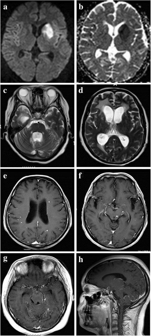
Fig. 1.

MR presentations of patients with tuberculous meningitis. a-bTBM patient with acute ischemic stroke in basal ganglia (hyperintense in DWI and hypointense in ADC); c-d TBM patient with hydrocephalus; e-f:TBM patient with multiple tuberculomas; g-h TBM patient with basal and cerebellar meningeal enhancement
