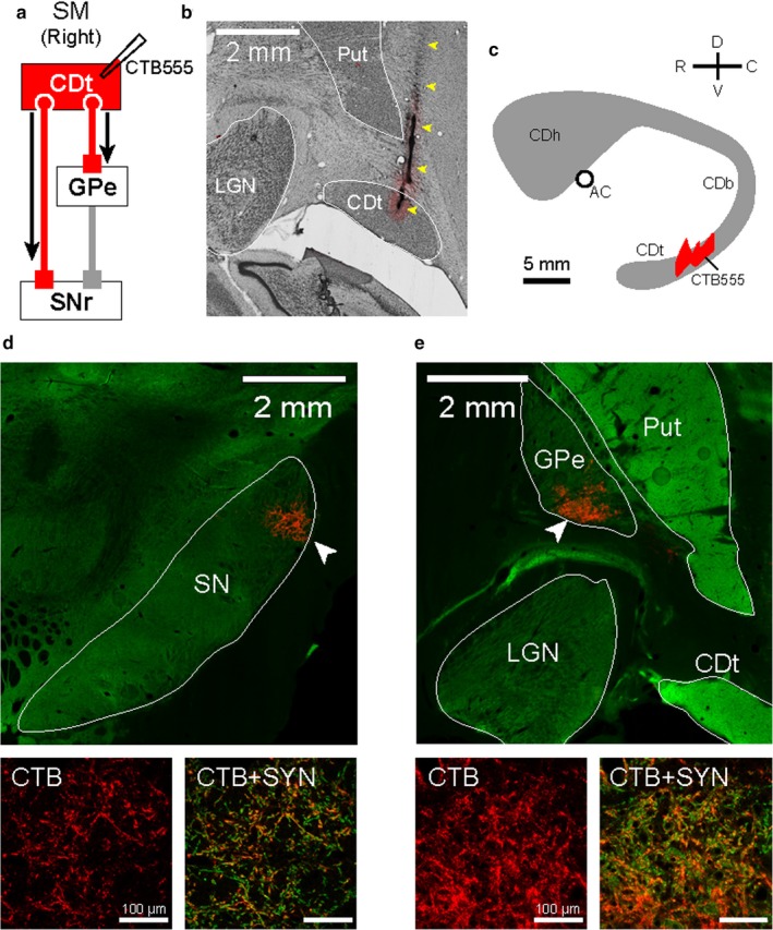
Figure 1.

CDt projected to the caudal parts of SNr and GPe. (a) Scheme of injection site in CDt of monkey SM. CTB555 was injected in CDt to identify its projection regions in the direct and indirect pathways. (b) CTB555 injection site in CDt. Injectrode track is indicated by yellow arrowheads. (c) Schematic injection locations at sagittal plane. CTB555 was injected in the middle region of CDt (see more details in the previous paper Kim et al., 2014). (d) CDt projection site in SNr which is shown by CTB signals (red). This was, localized in the caudal‐dorsal‐lateral part of SNr (cdlSNr; white arrowhead in top panel). CTB signals in cdlSNr (red in left‐bottom panel) were co‐localized with synaptophysin (SYN), an axon terminal marker (orange in right‐bottom panel). (e) CDt projection site in cvGPe. Axon terminals from CDt were localized in the caudal‐ventral part of GPe (cvGPe; white arrowhead in top panel). CTB signals in cvGPe (red in left‐bottom panel) were also co‐localized with SYN (orange in right‐bottom panel).
