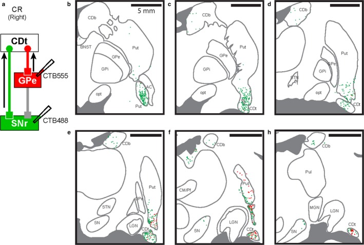
Figure 8.

cdlSNr‐ and cvGPe‐projecting neurons were spatially intermingled in CDt and cvPut. (a) Scheme of injection sites in cdlSNr and cvGPe of monkey CR. CTB488 and CTB555 were injected in cdlSNr (green) and cvGPe (red), respectively. (b‐g) Spatially intermingled projection neurons. Six coronal brain slices (P4, P6, P8, P10, P12 and P14) showed CTB488‐positive (green dots) and CTB555‐positive (red dots) neurons in the rostral‐caudal axis. cdlSNr‐ and cvGPe‐projecting neurons were spatially intermingled in cvPut and CDt (e‐g). Series of six slices from rostral to caudal at 2 mm intervals.
