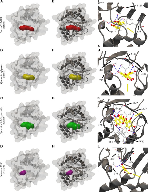
FIGURE 6.
Molecular docking of 2UV0 structure of LasR protein of P. aeruginosa PAO1 with 3-oxo-C12-HSL, quercetin aglycone, quercetin 3-β-D-glucoside and furanone C-30. (A–D) surface representation of 2UV0 structure of LasR protein of P. aeruginosa PAO1, (E–H) surface and backbone representations and (I–L) backbone representation with hydrogen bond between the amino acid residues and compounds evaluated. Gray surface representation, LasR protein; Red surface representation, 3-oxo-C12-HSL; Yellow surface representation, quercetin aglycone; Green surface representation, quercetin 3-β-D-glucoside; Purple surface representation, furanone C-30; Gray backbone representation, LasR protein; Black arrow indicates the binding site; Yellow arrow, 3-oxo-C12-HSL or quercetin aglycone or quercetin 3-β-D-glucoside or furanone C-30; Blue dashed line, hydrogen bond.

