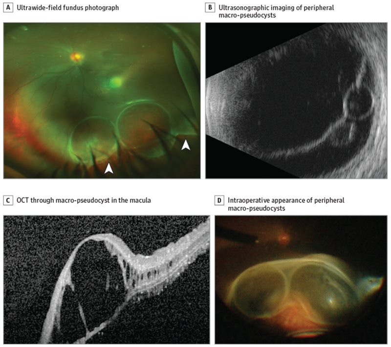
Figure 1. Multimodal Imaging.

A, Ultrawide-field fundus photograph showing a retinal tear (arrowheads), peripheral retinal macropseudocysts, and an inferior retinal detachment involving the macula. B, Ultrasonographic imaging of the peripheral retinal macro-pseudocysts within the detached retina. C, Optical coherence tomography (OCT) through the macular macro-pseudocyst showing splitting of the retinal layers. D, Intraoperative appearance ofthe peripheral retinal macro-pseudocysts on scleral depression.
