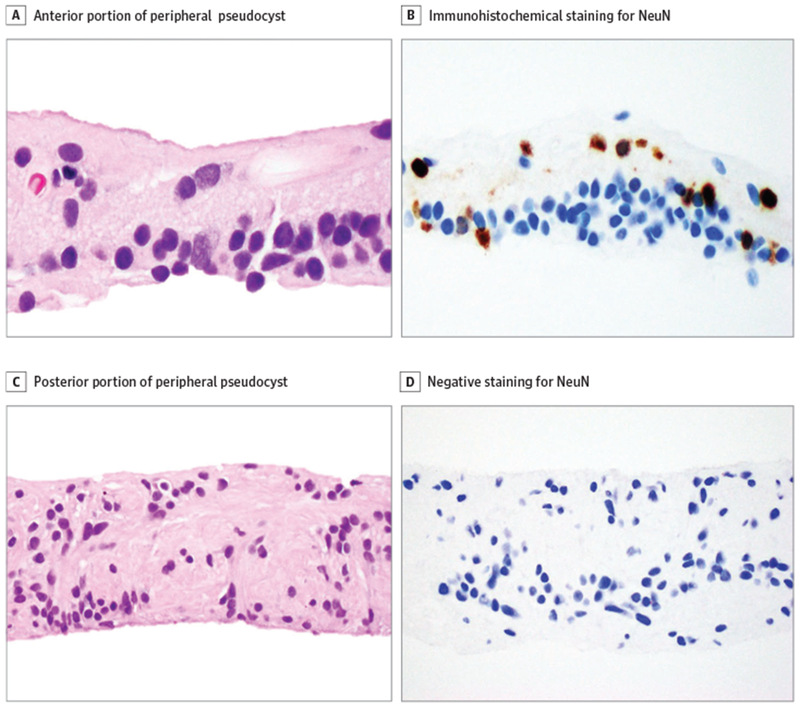
Figure 2. Histologic and Immunohistochemistry Findings.

A, Anterior portion of the peripheral pseudocyst. B, Immunohistochemical staining for NeuN was positive, highlighting ganglion cells and scattered cells of the inner nuclear layer. C, Posterior portion of the peripheral pseudocyst, showing disorganized glioneuronal tissue. D, Posterior portion of the peripheral pseudocyst, showing negative staining for NeuN. All parts, immunohistochemical stain, original magnification x400.
