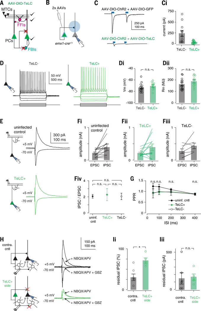
Fig. 4. TeLC expression selectively abolishes recurrent excitation.
(A) Schematic of circuit changes after TeLC expression in PCx principal cells. (B) Focal coinfection in PCx with ChR2 and either GFP or TeLC-GFP, followed by whole-cell recordings from uninfected cells. Light-evoked synaptic responses are abolished by TeLC. Example light-evoked response from non-ChR2–expressing neurons in (top) GFP- or (bottom) TeLC-GFP–infected PCx. i Light-evoked EPSC amplitudes in control and TeLC-expressing PCx (control: 239 ± 68 pA, n = 11 cells from two mice; TeLC 35 ± 10 pA, n = 12 cells from three mice; unpaired t test, P = 0.0133). (D) Example recordings from an (left) uninfected and (right) TeLC-infected neuron in the same slice in response to 50 pA current steps. (i) Resting membrane potentials (TeLC−, 73.4 ± 2.03 mV, n = 14 cells from three mice; TeLC+, 70.7 ± 2.01 mV, n = 11 cells from two mice; unpaired t test, P = 0.335) and (ii) input resistances (TeLC−, 162 ± 13.3 megohm; TeLC+, 188 ± 13.8 megohm; P = 0.188) were equivalent. (E) Synaptic inputs from OB are unaffected. Example recordings of EPSCs [membrane voltage (Vm), −70 mV] and disynaptic feedforward IPSCs (Vm, +5 mV) evoked by means of electrical stimulation of the lateral olfactory tract (LOT) in (top) an uninfected control slice or (bottom) a TeLC-infected neuron. Both EPSCs and IPSCs were blocked by 2,3-dihydroxy-6-nitro-7-sulfamoylbenzo[f]quinoxaline (NBQX) (10 μM) and D,L-2-amino-5-phosphonovaleric acid (APV) (50 μM, not shown). (F) Summary of LOT-evoked EPSC and IPSC amplitudes from (i) uninfected control slices, (ii) TeLC+ neurons and (iii) TeLC− neurons in TeLC-infected slices. (iv) EPSC/IPSC ratios were equivalent in all conditions; P > 0.05, unpaired t tests. (G) LOT EPSC paired-pulse ratios were not significantly altered after TeLC expression. n.s., not significant. (H) Example recordings showing recruitment of FBI is impaired, whereas FBI is unaffected. EPSCs and IPSCs were evoked by electrical stimulation of layer 2/3 226 ± 17 μm from recorded cell. EPSCs and IPSCs were attenuated in TeLC-infected slices. Blocking glutamate receptors with NBQX and APV eliminates the disynaptic component of IPSCs, with the residual IPSC evoked through direct stimulation of FBIs. The residual IPSC was fully blocked by gabazine (GBZ) (10 μM). (I) Summary of residual IPSC amplitudes. (i) The fractional size of residual IPSCs after NBQX/APV was substantially smaller in TeLC-infected slices (control, n = 6 cells from three mice; TeLC, n = 6 cells from three mice; unpaired t test, P = 0.0055), but (ii) the amplitudes of residual IPSCs were equivalent (P = 0.957).

