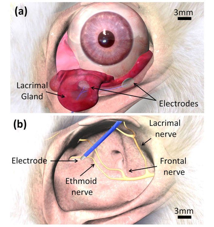
Figure 1.
Electrode placement in acute stimulation experiments.(a) The inferior lacrimal gland rests beneath the globe, behind the orbital rim. A bipolar electrode was implanted within the orbit, adjacent to the lacrimal gland. (b) The ethmoid nerve courses deep within the orbit, exiting posterior and nasal to the globe. A monopolar electrode was inserted through the caudal supraorbital incisure and advanced, behind the globe, toward the ethmoid nerve foramen. Blue color corresponds to insulation.

