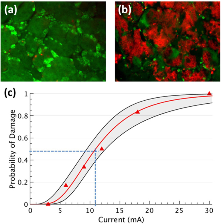
Figure 7.
Assessment of acute tissue damage. Probability of damage in excised pieces of lacrimal gland treated for 5 min with pulses of 1 ms duration at 30 Hz (2 mm diameter pipette electrode). (a) and (b) Show tissue stimulated with 3 and 30 mA, respectively. The color red marks cells with damaged cell membrane. (c) Probit analysis generated the best-fit curve and confidence intervals (68%, n = 6). Dashed lines indicate the threshold for acute lacrimal gland damage (50% probability of damage) to be 11 mA (3.5 μC mm−2 per pulse).

