
FIG. 1.
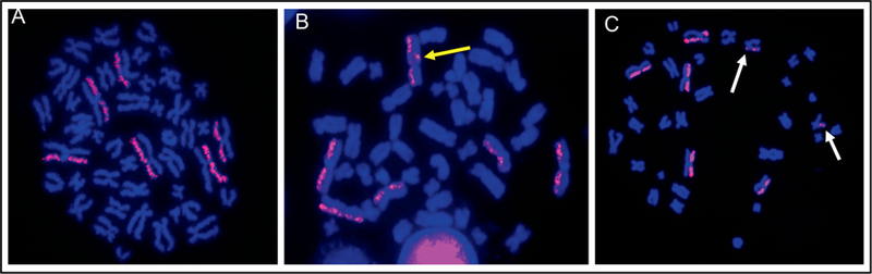
Directional genomic hybridization (dGH). Representative images of metaphase spreads labeled with dGH whole chromosome 1, 2 and 3 paints (red) and counter stained with DAPI (blue). Panel A: A normal metaphase spread free of any structural rearrangements. dGH chromosome paints uniformly label a single sister chromatid of a chromosome. Panel B: An inversion (double signal switch; yellow arrow) on chromosome 2. Panel C: Translocation involving chromosome 3 and a second, unpainted chromosome (white arrows).