
Figure 3.
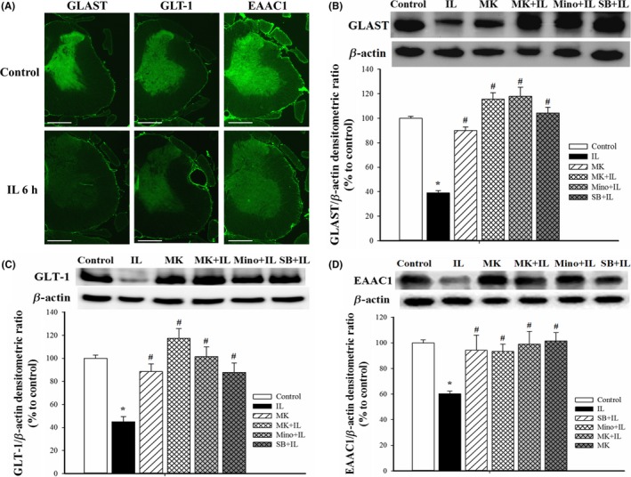
Effects of different treatments on the levels of glutamate transporters in SCDH. (A) Representative immunofluorescence images show the expression of GLAST, GLT‐1, and EAAC1 in the gray matter of spinal cord in controls (top row) and 6 h after IL‐1β injection (bottom row) (magnification: 25×; scale bar: 1000 μm). (B) Representative Western blotting results of GLAST (top), and quantification of these results in the five groups (bottom). Here and below, β‐actin was the loading control, quantification was performed with 4 rats per group, and values are expressed as means±SEMs. *P<.05 relative to controls; #P<.05 relative to rats given IL‐1β. (C). Representative Western blotting of GLT‐1 (top) and quantification of these results in the five groups (bottom). (D) Representative Western blotting of results of EAAC1 (top) and quantification of these results in the five groups (bottom)